That is where dental X-rays earn their keep. Long before a tooth stages a dramatic protest involving pain and a weekend emergency appointment, radiographs capture early evidence that something is off. They are less about confirming what can already be seen and more about exposing what prefers to stay hidden. Cavities may be the headline act, but they are far from the only story unfolding on those grayscale images.
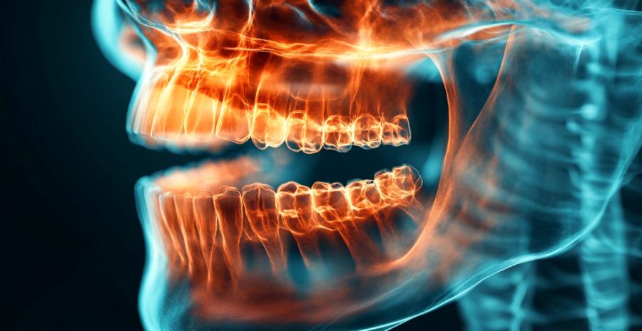
At a glance, an X-ray might resemble abstract art created by a machine with a limited color palette. To trained eyes, it is a detailed map of mineral density, anatomy, and subtle changes that would otherwise remain invisible. This is not guesswork or dental intuition; it is physics doing very practical work inside the mouth.
How dental X rays actually work
Dental radiography relies on controlled doses of electromagnetic radiation passing through the mouth. Dense structures like enamel and bone absorb more radiation and appear lighter, while softer tissues allow more rays to pass through and appear darker. This contrast is what creates the image.Modern systems use digital sensors instead of traditional film, which improves clarity and reduces exposure. The result is not only faster imaging but also the ability to enhance contrast and zoom in on suspicious areas. A shadow near a tooth root might look insignificant at first, but magnified, it can reveal early infection or bone changes.
There is something quietly humbling about the fact that a machine can detect microscopic changes long before nerves complain. Teeth, it turns out, are not always honest narrators of their own condition.
What shows up beyond cavities
Cavities are the most familiar finding, but they are only one item on a much longer list. Dental X-rays routinely reveal issues that have not yet earned symptoms or attention.- Bone loss associated with gum disease, often before gums look inflamed
- Infections at tooth roots that have not triggered pain
- Impacted teeth hiding below the surface, waiting patiently
- Cracks or fractures too fine to see during an exam
Why early detection matters more than drama
There is a persistent myth that dental problems announce themselves loudly and unmistakably. In reality, many of the most serious issues develop quietly. Bone loss, for example, can progress without discomfort until it reaches a point where options become limited.X-rays allow clinicians to intervene while problems are still manageable. This is not about creating extra work or inventing problems. It is about timing. A small infection treated early is usually straightforward. The same infection ignored for years tends to develop a personality and an invoice to match.
Radiation fears and the reality check
Mention X-rays outside a dental office and the imagination often leaps straight to hazmat suits and ominous warning signs. The reality is considerably less theatrical. Dental X-rays use extremely low levels of radiation, especially with modern digital systems.To put it in perspective, the exposure from a routine dental X-ray is often comparable to what the body absorbs during a short airplane flight or a day spent outdoors. No lead-lined bunker required. Protective measures such as lead aprons and thyroid collars further reduce exposure, largely out of an abundance of caution rather than necessity.
This is one of those areas where seriousness matters. Radiation safety is not treated casually in dentistry, and standards are guided by decades of research. The guiding principle is simple: use the lowest dose needed to get useful information, and only when there is a clear benefit. That balance is taken very seriously, even if the equipment itself looks deceptively friendly.
Why timing and frequency are individualized
Not everyone needs X-rays at the same interval. Someone with a history of frequent cavities or gum disease may benefit from more regular imaging, while others can go longer between scans. Age, oral health history, and current symptoms all factor into the decision.This individualized approach prevents both overuse and underuse. Too few images can allow problems to slip by unnoticed. Too many add no meaningful benefit. The goal is information that directly informs care, not a gallery of dental portraits collected for nostalgia.
There is also an efficiency factor. Having clear images on hand reduces guesswork, shortens appointments, and makes treatment planning far more precise. It is difficult to fix what cannot be seen, and even harder to explain it clearly without visual evidence.
Reading between the grayscale lines
Interpreting dental X-rays is a skill built on training and experience. What looks like a harmless shadow to one person might signal early bone changes to another. Context matters, comparison with previous images matters, and subtle shifts over time often matter most of all.This is where X-rays quietly shine. They are not dramatic on their own, but over years they tell a story of stability, improvement, or gradual decline. That long view helps guide smarter decisions and avoids reactionary treatment based on guesswork.
There is also something oddly reassuring about evidence. Seeing a problem clearly, even a small one, often reduces anxiety rather than increasing it. Uncertainty has a way of magnifying fear far more effectively than facts.
Getting to the root of the picture
Dental X-rays are not about hunting for bad news; they are about clarity. They reveal what eyes and mirrors cannot, long before discomfort demands attention. Used thoughtfully, they protect both oral health and peace of mind.Article kindly provided by soladentalspa.com